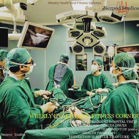
Weekly Health and Fitness Corner
Welcome to the "Weekly Health and Fitness Corner " where you get deep explanations and "candid discussions on critical "health and wellness issues" afflicting our society. There is an ever-increasing burden of "chronic diseases", primarily driven by a lack of common knowledge and the inability to ask questions. We need to know about these diseases in bits and reliable treatment procedures to follow up with. On this show, Healthcare professionals give a detailed breakdown of "over 5000+ human health problems. Enjoy the show. _NC Network. ® 2022
276
Jaundice - Prehepatic⧸Hepatic⧸Post Hepatic Caus...
"...jaundice is the yellow discoloration of the skin that is seen when bilirubin levels go above approximately 3 milligrams per deciliter but it can also be seen a particularly well in the sclera so first of all we need to know a little bit about bilirubin bilirubin is a breakdown product of him and is released from red blood cells when they are destroyed now bilirubin needs to get to the liver in order to be and the way it gets there is by initially being bound by albumin and then been transported to the liver via the blood it then gets taken up into the hepatic cells and is by the enzyme glucose urinal transfer is then secreted into the biliary system now this is the distinction between conjugated and unconjugated bilirubin the presence or absence of this glucose Iran sometimes this is referred to as soluble and insoluble bilirubin now direct and indirect bilirubin are often used as equivalence to conjugate it and unconjugated bilirubin but technically correct direct bilirubin includes conjugated bilirubin and Delta bilirubin which is the bilirubin bound to albumin that we mentioned earlier from there the conjugated bilirubin is present in the bayou and is secreted into the duodenum from the duodenum it travels through the small intestine up to the terminal ileum when most of the biological hepatic circulation but conjugated bilirubin is not reabsorbed it instead passes into the colon where the bacteria remove the gluco uronic acid that was added in the liver and forms urobilinogen which is colorless which is then oxidized into Starck or billion which gives feces it's brown color this is why in cases where the common bile duct is blocked you'll end up seeing pale stools right for anyone who's not been bored out of their mind by that bit will get into the causes of jaundice that you've probably heard split up into different categories these are pre hepatic hip-hop all post hypnotic jaundice which is also sometimes known as obstructive jaundice pre hepatic jaundice will have increased levels of unconjugated bilirubin because we are talking problem occurring before the bilirubin gets deliver these are going to be causes featuring excessive hemolysis so red blood cells being destroyed quicker than usual hemolytic anemia blood transfusions and hemolytic drugs hepatic causes are due to either having damaged hepato sites which is the case in hepatitis cirrhosis...""...jaundice is the yellow discoloration of the skin that is seen when bilirubin levels go above approximately 3 milligrams per deciliter but it can also be seen a particularly well in the sclera so first of all we need to know a little bit about bilirubin bilirubin is a breakdown product of him and is released from red blood cells when they are destroyed now bilirubin needs to get to the liver in order to be and the way it gets there is by initially being bound by albumin and then been transported to the liver via the blood it then gets taken up into the hepatic cells and is by the enzyme glucose urinal transfer is then secreted into the biliary system now this is the distinction between conjugated and unconjugated bilirubin the presence or absence of this glucose Iran sometimes this is referred to as soluble and insoluble bilirubin now direct and indirect bilirubin are often used as equivalence to conjugate it and unconjugated bilirubin but technically correct direct bilirubin includes conjugated bilirubin and Delta bilirubin which is the bilirubin bound to albumin that we mentioned earlier from there the conjugated bilirubin is present in the bayou and is secreted into the duodenum from the duodenum it travels through the small intestine up to the terminal ileum when most of the biological hepatic circulation but conjugated bilirubin is not reabsorbed it instead passes into the colon where the bacteria remove the gluco uronic acid that was added in the liver and forms urobilinogen which is colorless which is then oxidized into Starck or billion which gives feces it's brown color this is why in cases where the common bile duct is blocked you'll end up seeing pale stools right for anyone who's not been bored out of their mind by that bit will get into the causes of jaundice that you've probably heard split up into different categories these are pre hepatic hip-hop all post hypnotic jaundice which is also sometimes known as obstructive jaundice pre hepatic jaundice will have increased levels of unconjugated bilirubin because we are talking problem occurring before the bilirubin gets deliver these are going to be causes featuring excessive hemolysis so red blood cells being destroyed quicker than usual hemolytic anemia blood transfusions and hemolytic drugs hepatic causes are due to either having damaged hepato sites which is the case in hepatitis cirrhosis..."
6 min
277
Posterior Cerebral Artery Stroke Syndromes | PC...
"...stroke syndromes are collections of signs and symptoms resulting from Strokes in different regions of the brain or central nervous system in this video we will cover the stroke syndromes associated with Strokes in the territory of the posterior cerebral arteries they Supply the occipital lobe the inferior surface of the temporal lobes as well as some deeper structures such as the thalamus and the midbrain of the brainstem the posterior cerebral arteries arise from the distal end of the basilar artery which itself is formed from the vertebral arteries there is a connection with the anterior circulation via the posterior communicating artery our first syndrome is Alexia without a graph here which means the inability to read without the loss of writing here the affected regions are thus plenum of the corpus callosum and typically left occipital lobe the combination of these two areas being affected results in a pure word blindness meaning that the patient is able to write but is not able to read due to the involvement of the left occipital lobe contralateral homonymous hemianopia may also be present next is a similar syndrome the unilateral occipital stroke syndrome here as the name suggests the occipital lobe on one side side is affected and part of the inferior temporal lobe may also be involved in some instances the primary finding here is a contralateral homonymous hemianopia which may often be the only neurological deficit found we may see macular sparing due to the collateral blood supply to the macular region of the cortex in some instances and no Mia maybe present which is the inability to name in this case it's the inability T2 name colors and objects thirdly we have Anton syndrome also known as cortical blindness this occurs where there is an involvement of both occipital lobes either due to involvement of both posterior cerebral arteries or due to a lesion at the level of the distal basilar artery as a result there is visual loss however an interesting feature is that these patients may not be aware of the vision loss or we take this few our valued loyal listener about the best health and fitness podcast shows from the Nez pod Studios join us as we give you the best of the best health and wellness updates you can rely on for the treatment of chronic health problems classic functional medicine Back to Basics health tips and special updates..."
8 min
278
Portal Hypertension - Causes of Portal Hyperten...
"...portal hypertension refers to a higher than normal blood pressure in the portal system a normal range for this pressure is 5 to 10 millimeters of mercury portal hypertension can also be defined as a portal pressure more than 5 millimeters of mercury higher than the pressure in the inferior vena cava so the portal system refers to the portal vein which drains River and the main vessels that link to the portal vein and Superior mesenteric vein which comes from the small intestines the splenic vein which of course carries blood from the spleen the inferior mesenteric connects onto the splenic vein and carries blood from the large intestine but the gastric veins connect also on to the portal vein another thing to note is the umbilical vein which is normally obliterated and becomes the round ligament of the liver but if the pressure in the portal system gets high enough it can reopen varices are dilated veins that from an increased pressure in the portal system inside the liver you have structures known as sinusoids which are specialized capillaries within the liver the hepatocyte of the a separated from these sinusoids by a space known as the space of dese venous blood from the portal system mixes with arterial blood from the hepatic artery in the sign and then flows through into a central vein these Central veins collect together in the hepatic veins which takes blood into the inferior vena cava you can also see a which collects bile produced by the hepatocyte it's and takes it down towards the gallbladder the cell in red that you see inside the sinusoid is a cup for sale A specialized type of Mac the Scavenging and phagocytic activity the orange star shaped cells are important in Portal hypertension and cirrhosis these are hepatic stellate cells found in the space of these that are involved in fiberglass this and scaf formation in response to liver injury so what causes pressure in the portal system to increase the pathophysiology of portal hypertension ultimately comes down to blood being unable to pass smoothly from the portal circulation through the liver and into the inferior vena cava we typically divide the causes into pre hepatic intrahepatic post about it causes but remember that pre hepatic here means before the blood gets into the liver meaning causes in the portal vein itself well post hypnotic causes referred to causes after that two problems involving the inferior vena cava pre hepatic causes include portal vein thrombosis splenic vein thrombosis and arteriovenous malformation and splenomegaly intrahepatic causes include the most common cause cirrhosis which can come from alcohol abuse chronic viral hepatitis metabolic..."
8 min
279
Middle Cerebral Artery MCA Stroke Syndromes (Wi...
"...stroke syndromes are collections of signs and symptoms resulting from Strokes in different regions of the brain or central nervous system in this video we will cover the stroke syndromes associated with Strokes in of the middle cerebral artery IT supplies in most of the temporal lobe the anterolateral frontal lobe and the parietal lobe the middle cerebral artery comes off the internal carotid and is divided into segments the segments are the M1 or the horizontal segment which is the most proximal part and gives off a lenticular striate a tree which are deeper penetrating arteries that Supply the basal ganglia and surrounding region the M2 segment known as the Sylvian segment is next which typically includes a bifurcation into the superior and inferior segments em three segments a cortical supplying the cortex our first syndrome results from a stroke affecting the middle cerebral artery Superior division which normally supplies the lateral frontal lobe I'm the superior parietal lobes in this syndrome findings include contralateral weakness of the upper Limbs and the lower Limbs and a weakness of the contralateral lower face with a greater effect seen on the face and the Upper Limb rather than the lower limb this is because of the motor homunculus where we see that the areas of the cortex responsible for the legs are supplied more so by the anterior and so a less affected in Middle cerebral artery Strokes also remember that it is the lower part of the face that is affected because this only receives unilateral from the facial nerve while the upper two-thirds receives bilateral Innovation there is also often a Hemi sensory loss on the contralateral side that may affect the face or leg if the stroke involves the dominant hemisphere which is usually the left side then an expressive Aphasia it may be seen as Broca's area is found on the dominant side and is responsible for the production of speech if the stroke is on the non-dominant side then we see a contralateral any neglect where the patient may be unaware or unresponsive to stimulate on one side which may include not being able to recognize their own limbs next we have the middle cerebral artery inferior division..."
6 min
280
Signs of Cerebellar Dysfunction
"...Danish is a mnemonic to help you remember the cerebellar signs these are signs that indicate the presence of cerebellar lesions d stands for dysdiadochokinesia which is the inability to perform a rapid alternating movement such as flipping one hand against the palm of the other in a coordinated and controlled manner Desmet Rhea is a lack of coordination of movement involving the judgment of distance and this can be seen when asking the patient to reach a particular point with a finger a central Ataxia which literally means we take this few our valued loyal listener about the best health and fitness podcast shows from the Nez pod Studios join us as we give you the best of the best health and wellness updates you can rely on for the treatment of chronic health problems classic functional medicine Back to Basics health tips and special updates from the best doctors in the United States of America check out this health and wellness podcast shows explore Health talk weekly healthy lifestyle matters excellent Health digest healthy and free daily and weekly health and fitness Corner also check out nasty boy CC the truest story never told Fiction podcast for that real-life on-the-go experience with the 27 year old Golden Boy Who made our he tells us about his story as it happens in real time and in real life it's nasty boy CC the truest story never told go get a load of that happiness because happiness is healthy as we know it join us every week continue to provide you the best of health and fitness Wellness updates from around the globe enjoy the show means without coordination and this can affect not only the legs but also the arms trunk and other muscles in the body the most common manifestation we think of in this instance is an unsteady gait it can also be highlighted in the upper limbs by asking the patient to abduct their arms out to the side and then pressing down again and then Letting Go you may then see rebounding with a limb rebounds up Beyond its original position n is for nystagmus which are involuntary uncontrollable I move it can be elicited by asking the patient to move their gaze to the extremities and can have various characteristics including the speed rotation and Direction meaning either 200 vertical beating as well as to the left or to the right I is for intention tremor which is a Tremor that is worsened or exaggerated by movement or when the need for precise movement increases s stands for speech which may be slurred inappropriate or slow while he h stands for hypotonia..."
3 min
281
Post Stroke Complications - The most common com...
"...following a stroke there are several complications to be aware of the patient may have brain edema or increased intracranial pressure which can have a detrimental effect on the outcome of that patient this occurs due to Cellular swelling breakdown of the blood-brain barrier leaking of cerebrospinal fluid from the ependymal lining as well as from cellular debris causing an increased osmolality in that affected region this then causes movement of water into the affected space symptoms include headache dizziness nausea and vomiting and you may have signs such as papilledema and a gradual loss of consciousness because of the increased pressure there is also a risk of obstructive hydrocephalus who Nation Mannitol was previously used to reduce intracranial pressure in these patients however a recent study has shown that this may even increase the in-hospital mortality other potential therapies include hyperventilation and diuretics and some sources also specify steroids although the evidence is contradictory surgery involving a craniotomy with a skull is temporarily lifted can also help to reduce the intracranial pressure ischemic Strokes can give rise to a hemorrhagic transformation especially after thrombolytic treatment in the case of hemorrhagic stroke secondary to a subarachnoid hemorrhage the maybe vasospasm following the initial bleed which can then generate infarction which is essentially a secondary stroke to counter this a calcium channel blocker such as numata peen may be given and in the acute setting the patient may be given a cute volume expansion and vasopressin has in an attempt to increase cerebral blood flow infections are another complication of stroke pneumonia can be due to post stroke paralysis with a patient is not able to move well and can suffer atelectasis which is a collapse of part along this generates an environment that Mike I'm growing leading to pneumonia additionally due to swallowing difficulty the patient may be more prone to aspiration and therefore aspiration pneumonia urinary tract infections are also common especially in patients who have an indwelling catheter placed this is because patients with stroke have an increased risk from suppression bladder dysfunction and of course the use of the catheter itself Additionally the fever and systemic inflammatory response associated with the urinary tract infection May impair stroke recovery..."
5 min
282
Seizures | Generalized vs Focal Seizures | Caus...
9 min
283
Descending Tracts of the Spinal Cord
...the descending Pathways carry motor signals down the spinal cord and a generally divided into pyramidal or extrapyramidal tracts the pyramidal fibers travel through the medullary pyramids of the medulla oblongata which is why they are termed pyramidal these fibers originate in the cerebral cortex and are responsible for the voluntary control of the muscles of the body and the face there are two different tracks that make up the pyramidal tracts these are the corticospinal and corticobulbar tract the corticospinal tract responsible for the control of the body the cell bodies are found within the cerebral cortex with axons converging and passing through the internal capsule followed by the crus cerebri in the midbrain the pons and subsequently arriving into the medulla oblongata at this level around 75% the fibers will it to the other side of the spinal cord and continue down to sign ups with a lower motor neuron in the ventral horns at each level these fibers are known as the lateral corticospinal the other 25% of the fibers that remain on the ipsilateral side will continue as the anterior corticospinal tract and they will continue down ipsilateral e until the cervical and highest thoracic levels where they will decussate and sign ups with their motor neurons as for the corticobulbar tract this is the tract responsible for the voluntary control of the head the face and the neck similarly to the corticospinal tract they originated from the cerebral cortex and pass through the internal capsule however rather than descend into the spinal cord these neurons have their axons sign up sing to the lower motor neurons in the brain stem specifically on to the cranial nerve nuclei...
9 min
284
Syncope - What is Syncope?
"...the definition of syncope is a reversible loss of consciousness that occurs due to an inadequate blood flow to the brain usually their fast onset short duration and a spontaneous recovery so first things first what's the difference between a transient loss of consciousness and sinker beep well a syncope is a form of transient loss of consciousness which can be divided into loss of consciousness due to head trauma or non-traumatic causes under which comes along with syncope do other non-traumatic causes of a transient loss of consciousness include epileptic seizures psychogenic causes and rear causes like the problem clinically with syncope is Discerning the really serious and by that I mean potentially lethal causes of syncope so the causes are divided into Exxon knurled mediated syncope orthostatic hypotension and cardiac causes under reflex mediated we have vasovagal syncope which can be due to author's death that happens when the patient experiences fear phobias such as seeing blood or even pain next we have situational underneath reflex mediated syncope which includes losing Consciousness after death coughing finally for reflux syncope we have carotid sinus syndrome which is where the Carotid baroreceptors react too strongly to detecting an increased pressure leading to an excessive drop in blood pressure and therefore syncope an example is when people put their Ties on too tightly we take this few seconds off to inform you are valued loyal listener about the best health and fitness podcast shows from the Nez pod Studios join us as we give you the best of the best health and wellness updates you can rely on for the treatment of chronic classic functional medicine Back to Basics health tips and special updates from the best doctors in the United States of America check out this health and wellness podcast shows explore Health talk healthy lifestyle matters excellent Health digest healthy and free daily and last but not least weekly health and fitness Corner also check out nasty Boise see the truest story never told for that real life on the go experience with the 27 year old Golden Boy Who made our guest invite number one list he tells us about his story as it happens in real time and in real life it's nasty boy CC the truest story never told go get a load of that happiness because happiness is healthy as we know it join us every week as..."
6 min
285
Understanding Parkinson's Disease (Including Di...
"...Parkinson's disease is a neurodegenerative condition affecting dopaminergic neurons in the brain leading to Associated motor symptoms neurodegeneration means a progressive irreversible loss of neurons and in Parkinson's disease it is mostly the dopamine-producing neurons in the substantia nigra that's a lost for us to move signals are generated by the cerebral cortex that then pass through the motor pathways and lead to with our muscles and ultimately movement however this process needs to be regulated which is primarily the job of the basal ganglia the substantia nigra is a part of the basal ganglia which are a group of nuclei in the brain all normally there are two main Pathways the direct and indirect pathway the direct pathway is an excitatory pathway that facilitates movement the motor cortex sends excitatory signals via glutamate to the striatum the striatum sends inhibitory signals to the Globus pallidus internus substantia nigra pars reticulata via Gaba these two both release Gaba which normally inhibits Thalamus so in this path there is inhibition of the inhibition on the thalamus giving an overall increased activity of the thalamus which promotes movement the indirect pathway is instead an inhibitory pathway that's purpose is to terminate movement in this case there is an inhibitory Gaba signal from the striatum to the Globus pallidus external Tunis the Globus pallidus external is normally inhibits the subthalamic nucleus which normally stimulates the Globus pallidus internal this means that the indirect pathway increases the inhibitory effect of the Globus pallidus internus on the thalamus and so there is less signal from the thalamus and so there is less move in Parkinson's there is a gradual degeneration of neurons within the substantia nigra pars compacta these neurons release dopamine to the striatum in the striatum there are direct pathway neurons that have D1 dopamine receptors and are excited by dopamine and indirect pathway neurons that have dopamine D2 receptors and that inhibited by dopamine overall this means dopamine released from the past compactor..."
12 min
286
How to manage Hemorrhagic Stroke - Intracerebra...
"...stroke is characterized by having poor blood flow to part of the brain leading to cell death they are grossly divided into ischemic and hemorrhagic with around 15 to 20 percent of Strokes being hemorrhagic a hemorrhagic stroke results from the rupture of a blood vessel leading to bleeding compared ischemic stroke that have a sudden occlusion of a blood vessel within hemorrhagic Strokes there are two main types intracerebral meaning bleeding within the brain itself which can be intraparenchymal Hemorrhage weather is bleeding within the brain tissue or an intraventricular Hemorrhage where there is bleeding within the ventricular system of the brain around one intracerebral hemorrhage into the ventricles intracerebral Hemorrhage is most commonly caused by hypertension and age-related cerebral amyloid angiopathy which is where deposition of Lloyd Peter peptide in the vessels leads to a weaker vessel structure which is then therefore more likely to bleed the other main type is a subarachnoid with the bleed occurs between the arachnoid Mater and the Pia Mater subarachnoid hemorrhages can be due to trauma or can be spontaneous in 85% of cases in taneous subarachnoid hemorrhage is caused by rupture of a cerebral aneurysm with the most common locations being the anterior communicating artery in 35 percent of cases internal carotid artery in 30% and middle cerebral artery in 22% in 30% of cases there are multiple aneurysms the remaining maybe caused by rupture of an arteriovenous malformation coagulopathy or extension of an intraparenchymal bleed note that both of these types of hemorrhagic I considered intracranial bleeds however other types of intracranial bleeds such as epidural and subdural hemorrhages are not considered hemorrhagic stroke we take this few seconds off to inform you are valued loyal listener about the best health and fitness podcast shows from the Nez pod Studios join us as we give you the best of the best health and wellness updates you can rely on for the treatment of chronic classic functional medicine Back to Basics health tips and special updates from the best doctors in the United States of America check out this health and wellness podcast shows explore Health talk healthy lifestyle matters excellent Health digest healthy and free daily and last but not least weekly health and fitness Corner also check out nasty Boise see the truest story never told Fiction podcast for that real life on the go experience with the 27 year old Golden boy..."
8 min
287
Hepatorenal Syndrome Pathophysiology
"Hepatorenal syndrome is a life-threatening condition characterized by rapidly Progressive kidney failure that is seen in people with Advanced liver disease the prognosis is very Bleak and is usually fatal without the transplant there are two types type 1 has a median survival of two weeks and features a rapidly increasing creatinine level type 1 happens commonly in taneous bacterial peritonitis type 2 is a slightly more moderate form with a median survival of 10 weeks and a steadier creatinine here patients typically have a site that is resistant to DirectX approximately 18% of cirrhotic patients who have ascites will develop hepatorenal syndrome within one year so what exactly that makes this condition so bad the Hallmark is a renal basic constriction in the setting of vasodilation in splanchnic breasts those are the intestines spleen liver and pancreas the main branches of the aorta that make it up at the Celiac artery as well as the superior and inferior mesenteric arteries the underfill theory is that as liver disease progresses and portal hypertension follows possibly also generating ascites there is a splash so dilation because of release of vasodilatory mediators like nitric oxide and prostaglandins this vasodilation leads to more blood being directed into the splanchnic vessels which ends up draining into the portal circulation which causes the juxtaglomerular apparatus of the kidney to activate the renin-angiotensin-aldosterone system leading to vasoconstriction and in particular in the kidneys but the splanchnic vasculature is resistant to vasoconstriction due to the production of local vasodilators like nitric oxide and so remains vasodilate it the cycle leading to this vicious cycle of the renal vasoconstriction and splanchnic vasodilation ultimately leading to renal failure the activation of the renin-angiotensin-aldosterone is thought to be an important step in the formation of ascites in patients with cirrhosis to the extent that ascites and hepatorenal syndrome may be considered a splanchnic vasodilation determined the resistance of ascites to diuretics as is seen in type 2 hepatorenal syndrome as well as the onset of kidney vasoconstriction that leads to the initial onset of hepatorenal syndrome it is important to remember that having a cirrhotic patient with an AK I does not mean that the patient has HRS they may have the Aki for other reasons and so hepatorenal..."
6 min
288
Diarrhea Causes, Organic vs Functional Diarrhea...
"diarrhea is defined as having stools that occur more than three times a day and a looser than usual specifically 200 grams 24 hours is considered diagnostic but as you can imagine trying to measure the weight of stools in everyone referring to diarrhea would be a mess literally so first off the call divide them into acute and chronic causes for start acute diarrhea is defined as lasting less than four weeks and is often caused by infections that they usually eat a viral or toxin-mediated typically they will resolve spontaneously I see cases lasting more than four weeks need to be divided into either organic or functional causes and the difference between them is that organic causes can be detected or Quantified through testing while functional causes cannot samples of organic causes include celiacs disease inflammatory bowel disease like ulcerative colitis or Crohn's disease we can also have bacterial and parasitic infections pain and of course intestinal neoplasms on the other hand functional causes of chronic diarrhea include irritable bowel syndrome lactose intolerance food alcohol how then can we begin to narrow down the causes when we encounter a patient with diarrhea let's first tackle how to distinguish functional from organic diarrhea firstly the duration of secondly the volume of organic diarrhea is usually larger than in functional next we look for the presence or absence of blood functional will never have blood while organic often does we can also ask about the naming organic doesn't have any specific pattern but may wake the patient up at night well functional is usually in the morning and will not wake the patient up this is also closely tied to stress where organic diarrhea doesn't have much Association to stress but functional diarrhea often coincides with we take this few seconds our valued loyal listener about the best health and fitness podcast shows from the Nez pod Studios join us as we give you the best of the best health and wellness updates you can rely on for the treatment of chronic classic functional medicine Back to Basics health tips and special updates from the best doctors in the United States of America check out this health and wellness podcast shows explore Health talk weekly healthy lifestyle matters excellent Health digest healthy and free daily and last but not least weekly Health and Fitness also check out nasty boy CC the truest story never told Fiction podcast for that real-life on-the-go experience with the 27 year old Golden Boy Who made our guest he tells us about his story as it happens in real time and in real life it's nasty boy CC the truest story never told go get a load of that happiness because happiness is healthy as we know it join us every week as we continue to provide you the best of health and fitness Wellness updates from around the globe enjoy the show"
6 min
289
Nephritic Syndrome
Nephrotic syndrome is a collection of signs and symptoms that result from damage to the kidneys and is often confused with nephritic syndrome in fatigue syndrome there is a substantial amount of protein being lost through the kidneys in the urine defined as more than 3.5 grams per day this leads to hypoalbuminemia a low level of albumin in the blood as albumin is the most abundant protein normally in the blood these two are defining features in nephrotic syndrome nephritic syndrome is different in that there is less proteinuria but also the presence of hematuria and red blood cell or white blood cell casts in the urine hypertension and oliguria which is a reduced urine output typically between 80 and 400 ml per day the unit of the kidney is the nephron and normally in the glomerulus of the Nephron there is a specialized membrane that forms the filter made up of a fenestrated endothelium the Maryland basement membrane and the foot processes of podocytes which are cells that wrap around the capillary giving this additional filtration layer all together these structures normally act as a filter and prevent large molecules from passing through into the Bowman's capsule and renal tubules they have a net negative charge which may mean that they can repel other negatively charged molecules albumin is negatively charged in the fatigue syndrome there is sufficient injury to these structures to change the permeability and allow albumin and other molecules to pass into the urine leading to hypoalbuminemia the signs and symptoms linked to hypoalbuminemia which includes peripheral edema and fluid overload particularly in children this can be evident as facial swelling but can also occur in adults particularly around the eyes fluid overload can mean weight gain peripheral edema and even the development of ascites or pleural effusion which can manifest as shortness of breath known as this near the reduction in the blood levels of albumin causes the oncotic pressure of the blood 24 meaning fluid will more readily leak into the surrounding tissues causing edema this is sensed as hypovolemia because less fluid is in the vessels which then triggers the renin-angiotensin-aldosterone system causing retention of salt and water this is known as the underfill hypothesis while another is the
11 min
290
Acute kidney injury
a sudden deterioration in the function of the kidneys is known as an acute kidney injury sometimes also called acute renal failure to measure this reduction in kidney function glomerular filtration rate or GFR is normally used which is a measurement of how well the kidneys are filtering the blood the functional unit of the kidney that actually does this is the Nephron made up of the glomerulus which is a modified capillary and as blood passes through it the waste is filtered into the Bowman's capsule GFR is the volume filtered through the glomerulus into the Bowman's capsule and through the Nephron in a given unit of time the filtrate passes along through the proximal convoluted tubule Loop of henle distal convoluted tubule and the distal collecting duct undergoing secretion and reabsorptionas it travels ultimately it forms urine and collects into the renal pelvis and passes into the bladder through the ureter to directly measure this process is difficult we instead use creatinine clearance to estimate the GFR creatinine is a normal breakdown product of creatine released from muscle tissue and this is freely filtered by the glomerulus and is not reabsorbed so fits the kind of substance we need however creatinine is secreted into the filtrate by the peritubular capillaries so it does tend to overestimate the GFR slightly the cockcroft and Gault formula is a famous formula used to estimate the creatinine clearance and therefore GF are taking into account age Mass gender and the serum creatinine as it travels ultimately it forms urine and collects into the renal pelvis and passes into the bladder through the ureter to directly measure this process is difficult we instead use creatinine clearance to estimate the GFR creatinine is a normal breakdown product of creatine released from muscle tissue and this is freely filtered by the glomerulus and is not reabsorbed so fits the kind of substance we need however creatinine is secreted into the filtrate by the peritubular capillaries so it does tend to overestimate the GFR slightly the cockcroft and Gault formula is a famous formula used to estimate the creatinine clearance and therefore GF are taking into account age Mass gender and the serum creatinine
11 min
291
Polycystic kidney disease
polycystic kidney disease is a condition characterized by the development of multiple cysts within the renal tubules of the kidney and is the most common hereditary renal disease the normal functional unit of the kidney is the nephron made up of a glomerulus and Bowman's capsule proximal convoluted tubule Loop of henle distal convoluted tubule and the collecting duct blood filters through the glomerulus forming filtrate which then passes through the tubules undergoing see and reabsorption eventually forming urine and passing into the ureter and bladder overall there are around 1 million of these nephrons in each kid in polycystic kidney disease these tubules develop into cysts which become filled with fluid and can range in size from being microscopic to several centimeters in size the process can start even in utero meaning in the womb as the tubules become cysts they cannot carry the normal filtering function therefore the number of functioning nephrons begins to decrease this is worsened by the fact that as the cysts grow they compress nearby nephrons also making them dysfunctional initially this may not be seen to have any clinical impact as the remaining number of nephrons may increase there to maintain the glomerular filtration rate but as the disease progresses and more nephrons are affected the remaining ones cannot make up the difference overall renal function then deteriorates eventually reaching end-stage renal disease by definition the presence of structural injury makes polycystic kidney disease a form of chronic kidney disease with end-stage renal disease being defined as needing renal replacement therapy like dialysis or transplant or a GF far below 15 ml per minute there are two main types of polycystic kidney disease both caused by genetics the first is autosomal dominant with two main types one coming from mutations in P KD 1 on a chromosome 16 which codes for the protein polycystic in this is affected in around 85% of cases and is involved in cell to cell or cell Matrix interaction cell cycle regulation and calcium transport the second coming from mutations in PKD to on chromosome for coding for Polly system to which codes for
9 min
292
Chronic kidney disease
patients are 5 to 10 times more likely to die from cardiovascular disease than end-stage renal disease the risk of stroke has been found to increase as the GFR lowers around seven percent for every 10 ml per minute lost this may also be the result of CKD contributing to hypertension as well as being caused by it this can be due to activation of the renin-angiotensin-aldosterone system and can also be because as the disease progresses the kidney is less able to excrete leading to retention and subsequent fluid overload signs of fluid overload could be dismissed near due to pulmonary edema or a fusion and peripheral edema or ascites anemia is also common which contributes to disappear and can come from a reduction in erythropoietin from the
15 min
293
Nephritic Syndrome With Pathology
.nephritic syndrome is a collection of signs and symptoms that result from glomerulonephritis which is inflammation of the kidney specifically the glomerulus this could be remembered because it means inflammation rather than being a disease itself nephritic syndrome is the manifestation of an underlying disease that causes the inflammation and there are many of these causes it is easily confused with nephrotic syndrome which instead is a collection of signs and symptoms resulting from a high amount of protein being lost through the kidneys in the urine this leads to hypoalbuminemia meaning low levels of albumin in the blood these are the two defining features in nephrotic syndrome there is some overlap but nephritic syndrome is different in that the degree of proteinuria is lower and there is the dozens of hematuria meaning blood in the urine which could be microscopic or macroscopic red blood cell casts in the urine sterile / urea which is the presence of white blood cells without evidence of bacteria hypertension and oliguria which is a reduced urine output typically between 80 and 400 ml per day the functional unit of the kidney is the Nephron which includes the glomerulus and modified capillary surrounding this there are multiple layers which together form a filter these include a fenestrated endothelium the glomerular basement membrane and the foot processes of podocytes which are cells that wrap around the capillary in most cases of nephritic syndrome there is a trigger causing inflammation in this area
15 min
294
Tennis Elbow and Golfer's Elbow (Lateral & Medi...
"...tennis elbow and golfer's elbow a to similar conditions causing pain in the elbow and a medically know is lateral and medial epicondylitis respectively this means inflammation of the epicondyles which are bony protuberance has on the distal of the humerus the elbow is made up of the humerus articulated with the radius and ulna and allows for flexion and extension of the forearm however there are muscles that attach to the epicondyles that allow for wrist hand and finger movements like grasping and twisting and repetitive use of these muscles is thought to lead to micro attendance generating pain for this reason the conditions are known as enter Sapa these-- which means attachment point disease in act more recently it has been recommended that the conditions be referred to as tendinosis or epicondyle Alger rather than epicondylitis as histologically there is usually granulation tissue and a lack of traditional inflammatory cells suggesting that degeneration of the tendon maybe more an inflammation in the pathogenesis Motions like using handheld tools or even drawing are associated with the conditions and the names are slight misnomers is playing tennis can give you golfer's elbow and vice versa over all the movements causing the conditions do not necessarily need to be sports-related in some cases they can be triggered by a sudden contraction possibly from trauma elbow pain is the primary symptom in each lateral like this is 7 to 10 times more common and the pain is on the lateral or outer part of the elbow usually on the dominant hand it is generally worsened by extension of the wrist or fingers as the muscles responsible for these movements attached to the lateral epicondyle collectively known as the extensor tendons in particular the extensor carpi radialis brevis is commonly affected in lateral epicondylitis there is typically worsening of the pain with wrist extension against resistance when the is extended known as cozens test medial epicondylitis is less common but features pain on the medial or inner of the elbow it comes from tendinosis of the flexor and pronate attendance which originate from the medial epicondyle in particular the carpi radialis and the pronator teres and most commonly affected the pain is usually reproduced when the wrist is flexed and pronated against resistance well the is flexed called the reverse cozens test overall it is usually a gradual onset of pain that progressively get worse and can persist at night it also tends to be worsened by use of the forearm such as grasping and the pain may also be elicited by palpating several millimeters distal to the air condyles which corresponds with the location of the tendons people may also experience weaker grip strength the diagnosis is largely meaning no specific test or Imaging is required to make a diagnosis however Imaging like x-ray may be done to rule out arthritis of the elbow..."
7 min
295
Osgood Schlatter Disease (Tibial Tubercle Apoph...
"...Osgood-Schlatters disease also known as tibial tubercle apophysitis is a condition characterized by the anterior knee due to inflammation of the apophysis and apophysis is a normal developmental protuberance from a bone that provides an insertion site for tendons in this case the patellar tendon on to the tibial tubercle it primarily affects children and adolescents because by adulthood the apophysis will fuse with the rest of the tibia it is weaker than 10 and so is prone to injury Osgood-Schlatters disease is a stress or traction injury that occurs due to repetitive Force being applied through the strong patellar tendon on to the tibial tubercle typically from movements involving leg extension such as running or jumping but also during growth spurts the force can lead to small avulsion fractures of the tibial tubercle apophysis generating and pain and in rare cases a complete avulsion of the tibial tubercle knee pain is the most common symptom which is on the anterior portion of the knee just below the patella in most cases it is unilateral but both sides are affected in around 30 percent of cases in general it has an Insidious gradual onset with no trauma and may initially be episodic but go on to become continuous the tibial tubercle itself can be tender to touch and may have some Associated enlargement which can be felt as a prominent lump males in particular are up to seven times more likely to be affected..."
5 min
296
Ankle Fractures
"...ankle fractures a common injuries involving a break in one or more of the bones that make up the ankle joint there are three bones that make up the ankle the tibia fibula and tailless specifically the medial and posterior malleolus by the and the lateral malleolus of the fibula there is an additional joint between the tibia and fibula called the tibiofibular syndesmosis this is a fibrous structure made up of the anterior and posterior inferior tibiofibular ligament and the interosseous membrane the ankle is a hinge primarily allowing supplanter and dorsiflexion and is at its least stable in plantarflexion it is described as a mortise and Tenon joint which are essentially a hole and the that fits that hole in this case the space formed by the tibia and fibula and the projection which is the Dome of the talus bone ligaments helped provide more stabilization there is the medial or deltoid ligament coming from the medial malleolus which branches into four ligaments and is mostly involved in preventing / e-version while the lateral ligament comes from the lateral malleolus and is made up of three ligaments with the main function of preventing over inversion of the foot in 70% of cases there is an isolated fracture of either the medial or lateral malleolus if both the fractured this is termed a bye the fracture seen in twenty percent of cases and if the posterior malleolus is also fractured this is called a try malleolus fracture but this is only the case in fewer than ten percent of patients in roughly 10% of cases the syndesmosis is also injured there are multiple classifications of ankle weather is used in the lateral malleolus fractures based on the location of the fracture relative to the syndesmosis generally with more proximal fractures indicating a higher risk of instability a is below the syndesmosis B is at its level and see is proximal to it the log handsome classification is more detailed considering also the position of the anchor during injury and the forces sustained during it other types of fractures include Amazon nerve injury where there is a proximal fibula fracture with syndesmosis and deltoid ligament injury this is an unstable injury that will need surgical intervention this is..."
9 min
297
Shoulder Dislocation (Glenohumeral Joint Disloc...
...arm in an anterior dislocation due to the posterolateral portion of the humeral head being compressed against the anterior part of the glenoid labrum they can be compression fractures on the humerus known as Hill Sachs lesions they are closely linked with bankart lesions which is a rupture in the glenoid labrum these can also be accompanied by avulsion fractures which are then termed bony bankart lesions the arm is typically held in external rotation with some abduction and all movements are painful there is a loss of the normal Contour of the deltoid and the acromion can be particularly prominent posterior dislocation 's make up only two to four percent of all shoulder dislocations and occur due to the head of the humerus being forced posteriorly while...
9 min
298
Stress Response - Health FAQs with Dr Gates
"...for those of you who have not tuned in to us we run essentially a chronic pain practice board Gates is board certified functional neurologist I'm a board-certified functional medicine practitioner we melded those two disciplines into a model of care to address a substantial portion of chronic conditions chronic Jesus and and although we cure few we're able to help most get under some some control quite considerable control and be able to manage their case and the properly selected and the properly cite the patient the reason we cure for you is because pretty much everybody walks in here ends up having an autoimmune problem and they also usually walk in here under a chronic stress with a chronic stress response usually we caught a sympathetic dominance or fight-flight system is engaged and it doesn't tend to let go these things can lead to anxiety Sia T panic attacks depression things of that nature and I lead off with that to say one of my one of my one of my points with people when they come in and I address this with them is my observation based on watching what happens with the brain rehab exercises that we do in certain other types of supplementation is that these are not necessarily personality disorders their poor frontal lobe this sort of and there's a lot of things that add to causing your frontal lobe to not work properly poor poor lack of blood supply to your Bruno lobe from anemias be your frontal lobe needs oxygen inflammation which are probably going to talk about a little bit blood sugar stress hormones and and so but lately we've been seeing last couple of years maybe we've been even seeing little kids come in we're stressed out of their minds and like two or three years old and say what's going on with that and then other people who've had just this kind of stress responses that seemed to be like even a little bit over a top for what we see and and dr. Gates is going to share with you some of the things that he's come across in that in the literature that we've been implementing and what are our Protocols are and kind of giving you the understanding that I know stress hormones now can cross the placenta from the stressed mother into the young person but there's even more to it than that our genetic components that need to be considered if they're there and that oftentimes respond well to the types of things that can be done wow without drugs frankly so yeah we treat a lot of patients with fibromyalgia in our clinic at least we have and that's actually started off with yeah that's kind of how we got into depression because a lot of patients were coming in with their fibromyalgia and they didn't want to take medications for the depression so they said well can you handle what could you do so that started us on this quest in this journey of..."
9 min
299
Hashimoto and Plastics - Health FAQs with Dr Gates
.so we're going to talk about Hashimoto and Plastics today Hashimoto's is a subject that we've been long associated with frankly probably for most people are associated with and we're taking quite a bit of use for even diagnosing people at and so and getting heat on it and the point from that is that Hashimoto is the understanding of what's going on with Hashimoto's is the number of things it's related to the things that can create a Hashimoto's isn't it is an immune attack on your thyroid primarily and so you're always looking for the things that are creating the immune attack and and and it can be a multitude of things and I was just mentioning to dr. Gates we're talking about Plastics that I remember reading about that at like 10 years ago but wondering is actually something that has something to do with it because back then it was I was everything was Metals was right it was it was is there anything you could think of the alternative groups were putting out as yet avoid all of this which is basically everything in life it seemed like but it turns out that Plastics do create a negative effect on the condition of Hashimoto's and so we're going to discuss that this probably fairly briefly it's not a long topic I would assume and so and if it's not brief then that's fine too so so Plastics as the famous gentleman in the Dustin Hoffman movie from many years ago The Graduate said to Dustin Hoffman when he had him in his graduate courses okay so I'm 65 days going to be people more my age The Graduate it was the famous line for the decade
8 min
300
Immune System - Health FAQs With Dr. Gates
"...and today we're going to talk about a concept that has time so I think been controversial I think at times been considered theoretical when I first got into functional medicine years ago is actually a prominent Conrad of the immune system and because what we started to realize was in chronic pain we have a chronic pain practice for those of you who haven't watched us before and we treat just about anything that won't go away and we've combined two disciplines functional neurology and functional medicine to address fibromyalgia and periphery often chronic fatigue and chronic got problems chronic thyroid problems and dizziness vertigo balance things that won't go away people get things they go away B they don't go away you need to know why and the immune system was an obvious first place to start I think for those in the field at that time that there must be something with the immune that's causing the problem there's a concept called th1 th2 shifts th1 th2 balance I've drawn the little seesaw picture of it and I think a thousand times and try and explain to people and and and I think it's I don't I think dr. Gates is going to explain to me today whether it's getting more credibility or just something that you're using more because you see it's a valuable tool because it is affecting people frequently will talk about that Gates does all the treatment now and he's always researching and frequently will tell talk to me about how this was a that this was a shift during pregnancy this is the mystical why do I get pregnant and then I feel good and then all sudden I don't after I deliver the child it's this is the if you have allergies you have a shift more towards one side than the other so and certain autoimmune problems have a certain quality..."
16 min